
Virtual ResusX:2026 Is LIVE (Early-Bird • Save 50%)
The wait is over!
Today you can grab your virtual ticket to ResusX:2026 — and join us from anywhere in the world.
Whether you can’t travel or simply prefer to learn from home, this is your opportunity to experience the energy, insight, and high-yield education that makes ResusX unforgettable.
With your Virtual Ticket, you’ll get:
-
🎥 Access to powerful, high-impact resuscitation talks
-
🧠 Practical clinical pearls you can apply immediately
-
🌎 Earn CME / CEU from the comfort of your own home
-
🔥 The same bold, engaging ResusX experience — streamed to you
ResusX isn’t passive learning.
ResusX is immersive. Practical. Real-world resuscitation education.
Don't sleep on this 50% discount...these coupon codes will go quick so don't wait!
I can’t wait to have you join us for ResusX:2026.
Haney (@CriticalCareNow)
Welcome To ResusNation #148
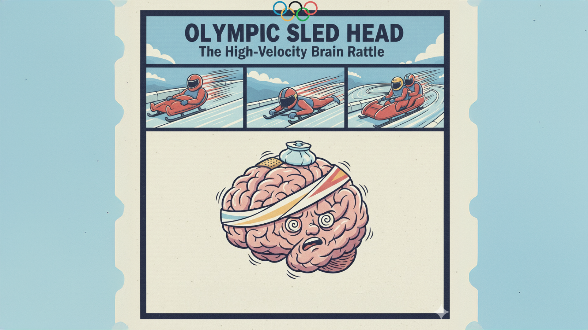
Sled Head: When Your Brain Becomes a Human Pinball
Imagine voluntarily signing up to be a human bullet, greasing yourself up to slide down an icy tube at 90 mph while pulling 5Gs on every curve. That’s the vibe of Olympic sliding sports (luge, skeleton, and bobsled), but the real party starts in the skull with a phenomenon lovingly known as "Sled Head." It’s essentially what happens when your brain decides to play a high-stakes game of Pong against your own cranium. Every vibration from the track and every micro-impact isn't just a thrill—it’s a repetitive sub-concussive rattling that leaves athletes dealing with a cocktail of brain fog, chronic headaches, and the kind of psychological fatigue that makes a 30-hour call shift look like a spa day. We’re not talking about one big "lights out" hit; it’s the cumulative "death by a thousand vibrations" that turns an elite athlete's neurological profile into a chaotic mess of vestibular dysfunction.
The real kicker is that the culture of these sports is basically "rub some ice on it and go again," which is a bold strategy when your white matter is screaming for a ceasefire. Because these athletes are effectively living in a high-speed paint shaker, the symptoms often mimic a slow-motion concussion that never actually gets the chance to heal. Researchers are now looking at the long-term neurovascular fallout, realizing that the "post-run daze" isn't just adrenaline—it’s the brain’s way of asking why you’ve chosen a career path that involves literal gravitational assault. It’s a fascinating, terrifying look at how much mechanical stress the human nervous system can take before it starts sending out the "Internal Server Error" signals, proving once again that gravity is a cruel mistress with zero respect for your Hippocratic Oath or your pre-frontal cortex.

Why We Stopped Cooling Post-ROSC
The field moved from 33°C (active cooling) to 36°C after the original TTM trial (Nielsen et al., 2013) showed no difference in outcomes between the two targets. This was already a significant shift, as 33°C came with real costs — arrhythmias, hypotension, coagulopathies, and infection risk.
Then TTM2 (Dankiewicz et al., 2021) was the game-changer. This large RCT compared active cooling to 33°C versus normothermia with strict fever prevention, and found no survival or neurological benefit to active hypothermia. The key insight was reframing the question entirely: the harm isn't from not cooling — it's from allowing fever.
The current paradigm: The goal post-ROSC is now aggressive fever prevention (generally keeping temperature below 37.5°C) rather than active cooling. This is now reflected in major guidelines including AHA and ERC/ESICM. It's a much more manageable target that avoids the complications of induced hypothermia while still protecting the vulnerable post-arrest brain. The underlying physiology makes sense — the ischemia-reperfusion injured brain is highly sensitive to hyperthermia, which worsens cerebral metabolic demand, excitotoxicity, and cerebral edema.
Watch the full video here and leave a comment.
Don't forget to like and follow my IG, TikTok, YT, Facebook or Linkedin accounts.


Why 30cc/kg is Killing Your Heart Patients
Managing cardiogenic shock requires a shift from idealistic protocols to a pragmatic, clinical focus on perfusion and volume status. The most critical error in the emergency setting is the reflexive administration of the CMS-mandated $30\text{ mL/kg}$ fluid bolus, which is designed for distributive (septic) shock but can be fatal for a failing heart. By categorizing patients based on their physical presentation—specifically looking for the "wet and cold" phenotype—clinicians can identify true cardiogenic shock early. Tools like Point of Care Ultrasound (POCUS) and the monitoring of $\text{SCV}\text{O}_2$ (Central Venous Oxygen Saturation) serve as vital surrogates for cardiac output; notably, an $\text{SCV}\text{O}_2$ below $50\%$ indicates a patient in extremis, while a $4\%$ rise following a small fluid challenge can safely identify fluid-responsive patients.
Understanding the trajectory of the disease is essential for triage, as mortality rates climb drastically from Stage A through Stage E of the SCAI shock criteria. While Stage B patients may appear "normal" with stable blood pressure, they carry a $34\%$ mortality rate, which jumps to $77\%$ in Stage E "extremis" patients who have suffered cardiac arrest. Ultimately, the pragmatic provider must recognize that while vasopressors and inotropes stabilize the patient, they do not "fix" the heart. The goal of medical management is to buy time for a multidisciplinary team—including interventional cardiologists and cardiac surgeons—to provide definitive mechanical or surgical intervention before the patient deteriorates past the point of no return.
Check out this video of Dr. Eddy Gutierrez from ResusX:2025 now!


One Size Doesn't Fit All In ARDS
Takotsubo syndrome (TTS) is no longer just a "broken heart" curiosity; this review highlights it as a complex, inflammatory state with significant morbidity and mortality risks that can equal those of acute myocardial infarction. While we have traditionally leaned on the catecholamine surge theory, the data now points to a central role for the NLRP3 inflammasome and a systemic "cytokine storm" (IL-6, IL-1B, TNF-α) that drives myocardial stunning and microvascular dysfunction. Essentially, this article reframes TTS as a neuro-cardiac-inflammatory syndrome where the intensity of the inflammatory response, specifically markers like the neutrophil-to-lymphocyte ratio (NLR), directly correlates with poor long-term outcomes and recurrence risk.
From a practice standpoint, this shifts our focus from simple supportive care to a more aggressive, multi-modality diagnostic and potentially disease-modifying approach. While we still treat the acute heart failure symptoms, the standard dogma of long-term beta-blocker therapy is being questioned, as large registries show no benefit in reducing recurrence. Instead, the impact here is on the horizon: we are moving toward using cardiac MRI (CMR) not just for diagnosis, but for assessing residual inflammation (T1/T2 mapping) to decide if a patient has truly recovered. If we see persistent edema or atypical patterns, we need to be more vigilant about complications like LV thrombus or recurrence.
My Takeaway Points
-
Inflammation as a Prognostic Tool: Don't ignore the basic white cell count; systemic inflammatory markers like high NLR or low lymphocyte-to-monocyte ratio (LMR) are independent predictors of long-term mortality in TTS.
-
The Beta-Blocker Myth: Be cautious with the assumption that beta-blockers prevent recurrence; observational data (GEIST and SWEDEHEART registries) suggests they may not lower recurrence risk, though they remain useful for acute sympathetic blunting and heart failure management.
-
CMR is the Gold Standard for Clearance: If a patient has persistent dyspnea or fatigue months later, it’s likely subclinical inflammation. Use CMR tissue characterization (T2 mapping) to look for ongoing edema, as this helps risk-stratify those at higher risk for recurrence.
-
Emerging Therapeutics: Watch for the results of the CIT-DZHK29 trial; we may soon be using short-term Cyclosporine A or IL-1 blockers (Anakinra) to dampen the acute inflammatory burst and improve myocardial recovery.
-
Pro-Tip (Patient Communication): When explaining the "broken heart" to a patient, avoid the term "benign." Tell them: "While your heart muscle isn't permanently blocked like a typical heart attack, it has experienced a 'short-circuit' and significant inflammation. We need to monitor you closely for the next 3 months—just like a standard heart attack—to make sure the 'swelling' in the heart muscle goes away completely."
Want to learn more? Read the full article "The Role of Inflammation in Takotsubo Syndrome: From Pathogenic Pathways to Imaging Insights and Therapeutic Perspectives" by Madaudo et al. in Current Cardiology Reports.
Ventilator Alarms: A Simple Way to Think When the Noise Starts
Ventilator alarms have a habit of going off when the room is already busy and the patient is already sick. The problem usually isn’t the alarm itself, but how quickly we slip into reflex mode: silence it, stare at the ventilator, and start changing settings without a clear plan. When an alarm goes off, slow things down just enough to apply a simple framework:
1. CONFIRM the problem is real. EtCO₂ lines kink or fill with moisture, patients cough, and alarm limits are sometimes set inappropriately, all of which can trigger alarms without meaningful physiologic change.
2. ASSESS the patient, not the machine. Look at chest rise, synchrony, work of breathing, blood pressure, and overall stability. If the patient looks unstable, it may be better to disconnect and bag while you regroup.
3. INVESTIGATE systematically, always working from the patient back to the ventilator. Listen for bilateral breath sounds and consider pneumothorax or right mainstem intubation. Check tube patency and suction early. This fixes more vent alarms than people realize. If those check out, then move to the circuit and finally the ventilator.
4. INTERVENE: make one intentional change based on your working diagnosis, rather than “turning knobs until it stops beeping.”
5. REASSESS the patient and waveform to see if it actually helped. Layering multiple adjustments without reassessment is where trouble starts.
Ventilator alarms aren’t emergencies by themselves. Having a structured way to respond helps keep small problems from compounding when the room is already loud.
-----------
Dr. Shawn Segeren is a Canada-based Emergency Medicine physician and founder of Dynamic Simulation, a CME-accredited clinical simulation program delivering interprofessional, critical care simulations in emergency departments across Ontario.
Connect with Dr. Segeren:
Watch the February Videos Now!
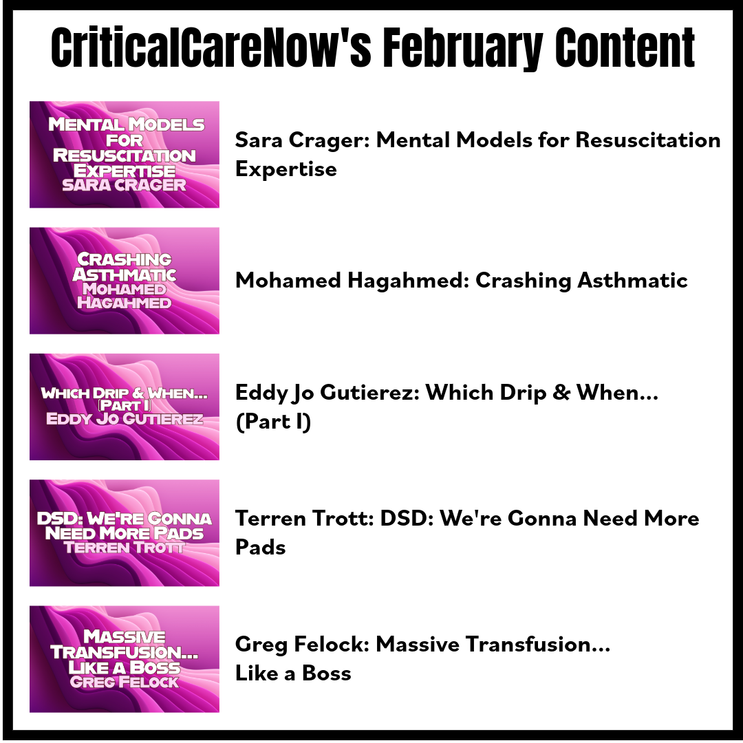
If you're an All-Access Member, you're in for some great content this month. We have FIVE videos hand-picked by our staff that are high-yield and our most highly watched. We're featuring
- Crager on "Mental Models for Resuscitation Expertise"
- Hagahmed on "Crashing Asthmatic"
- Gutierez on "Which Drip & When... (Part I)"
- Trott on "DSD: We're Gonna Need More Pads"
- Felock on "Massive Transfusion... Like a Boss"
Each month we bring you fresh new content from the best of the best in resuscitation. If you're an All-Access member, go watch these videos NOW! If you're not, then sign up here.


Responses