
Welcome to ResusNation #152


If You Can’t Handle Me at My Lapalus, You Don’t Deserve My PB
Cross-country skiing and biathlon are essentially organized torture sessions where your heart rate is redlining at 190 bpm while you're actively being flash-frozen. Because these athletes are basically human jet engines breathing in dry, sub-zero air, their respiratory tracts go into a full-blown panic. This leads to a phenomenon the pros call "ski lung," but the visual reality is much more chaotic: exercise-induced rhinorrhea and hypersalivation. Your body, in its infinite wisdom, tries to humidify that arctic air by pumping out massive amounts of fluid, which then hit the 20-mph headwind and immediately decide to vacate the premises via your nostrils and mouth. It’s a literal flood of secretions that would make an ICU nurse reach for the Yankauer, but instead of being suctioned, it just migrates.
Once that liquid gold makes its exit, physics takes over and things get truly gnarly. As the sweat, snot, and spit meet the freezing ambient temperature and the high-velocity airflow of a downhill descent, they undergo a phase change directly on the athlete's skin. We’re talking about a structural layering of frozen biological fluids that creates a literal "beard of ice" or the legendary "snotcicle." French skier Hugo Lapalus has basically become the patron saint of this aesthetic; the man finishes races looking like he’s wearing a crystal chandelier made of his own DNA. It’s the ultimate "forbidden frosting"—a salty, frozen testament to the fact that the human body is remarkably efficient at turning peak athletic performance into a biological slushie.

We're hosting a medical conference at a comedy club.
No, really.
ResusX 2026 lands May 18–20 in Philadelphia — not at a hotel ballroom with fluorescent lights and lukewarm coffee. At the Punch Line Comedy Club.
Here's why that matters:
Comedy clubs are engineered to hold attention. The stage is close. The seats are intimate. The room doesn't let you zone out — which means the 2pm session hits exactly like the 9am one.
And what's filling those sessions?
→ Live procedural demos. Not clips. Not diagrams. Live.
→ Expert debates that actually change how you think.
→ Interactive formats where you're the one making the calls.
→ High-momentum learning built for retention, not résumés.
→ Heavy content. Unforgettable room.
If you've been telling yourself you'll level up your resuscitation skills "soon" — this is what soon looks like.
→ Not an attending physician? You're still invited. Fill out this form to unlock a discount code built for non-attending clinicians.

Pain First, Sedation Second: The Case for Analgosedation in the ICU
ICU patients on sedation are often undertreated for pain, leading clinicians to reflexively escalate sedatives like propofol when the real issue is unaddressed analgesia. The SCCM guidelines address this directly through the principle of analgosedation — front-loading analgesia before initiating or escalating sedation. This makes physiological sense given how many sources of discomfort these patients face: endotracheal tubes, central lines, prolonged immobility, and frequent procedures. When pain is adequately treated first, the required sedative doses drop considerably.
This approach has meaningful clinical consequences beyond patient comfort. Undertreated pain masked by heavy sedation still causes physiological stress, and excessive sedation is independently associated with increased delirium in the ICU. Delirium, in turn, is linked to higher mortality, longer ventilator dependence, and extended ICU stays. Shifting to an analgesia-first model has been one of the more impactful cultural and practice changes in critical care in recent years, with real downstream effects on patient outcomes.
Watch the full video here and leave a comment.
Don't forget to like and follow my IG, TikTok, YT, Facebook or Linkedin accounts.


Modern emergency medicine is witnessing a paradigm shift in how we approach post-cardiac arrest care, specifically regarding the "reflex" trip to the cath lab. While coronary artery disease remains the primary cause of out-of-hospital cardiac arrest, recent landmark clinical trials like COACT (2019) and TOMAHAWK (2021) have challenged the traditional urgency of immediate intervention. These studies demonstrate that for patients without an obvious STEMI, an early, immediate coronary angiography strategy does not improve mortality or neurological outcomes compared to a delayed, selective approach. Essentially, once the initial anoxic brain injury has occurred, opening a non-occluded coronary artery immediately provides little systemic benefit, allowing clinicians to focus on high-quality supportive care first.
However, the "death of the post-arrest cath" is a strategic oversimplification. Data from these same trials reveal a significant crossover rate—nearly 20% of patients initially assigned to the delayed group still required emergent intervention due to clinical deterioration. Dr. Trott emphasizes that while we have moved away from cathing every shockable rhythm, there are three non-negotiable "red flags" that still demand immediate activation: STEMI, refractory arrhythmias, and profound cardiogenic shock. The goal is for clinicians is to move from a "cath everyone" mindset to a selective approach, identifying the specific patients where reperfusion actually prevents a re-arrest.
Check out this video of Dr. Terren Trott from ResusX:2025 now!


Why Withholding Contrast May Be Wrong for CKD Patients
Decades of fear surrounding iodinated contrast media and acute kidney injury (AKI) were largely built on outdated 1950s case reports using high-osmolar agents no longer in clinical use. A 2026 review in Kidney360 by Florens and Demiselle systematically challenges this paradigm, demonstrating that modern low- and isoosmolar contrast agents rarely cause true nephrotoxicity — even in high-risk populations with advanced CKD or active AKI. Early observational studies lacked proper control groups, meaning post-procedure creatinine rises were reflexively attributed to contrast despite confounders like hemodynamic instability or concurrent nephrotoxins. Propensity-matched data from over 53,000 CT patients found no difference in AKI rates between contrast and non-contrast scans, and landmark trials like PRESERVE and AMACING have since invalidated the two most common prophylactic strategies — sodium bicarbonate with N-acetylcysteine, and routine IV hydration — respectively.
The real danger, the authors argue, is renalism — the well-documented phenomenon of withholding clinically necessary imaging out of exaggerated nephrotoxicity fear. This leads to missed diagnoses, delays, and inferior alternatives. Chertow, et al. previously showed that CKD patients with MI denied coronary angiography on these grounds had measurably higher mortality. For the vast majority of patients, including those with mild-to-moderate CKD, the risk-benefit calculation strongly favors proceeding with indicated contrast imaging. Reasonable precautions — avoiding hypovolemia, minimizing contrast dose, and holding concurrent nephrotoxins — remain appropriate for an eGFR below 30 ml/min, but blanket avoidance is no longer defensible.
My Takeaway Points:
-
Finding - Propensity-matched studies including 53,000+ CT patients demonstrate no significant difference in AKI rates between contrast-exposed and unexposed patients, even among those with advanced CKD or active AKI on presentation.
-
Practice Impact - Routine prophylactic IV hydration and N-acetylcysteine are not supported by RCT evidence and should not delay or replace clinically indicated contrast imaging; individualized risk-benefit analysis should replace blanket contrast avoidance.
-
Population - High-risk patients including those with CKD (eGFR 30–59), active AKI, critically ill ICU patients, and elderly patients with comorbidities — historically the most contrast-restricted groups — showed minimal to no contrast-attributable renal harm in contemporary controlled analyses.
-
Limitation - Most supporting studies are propensity-matched retrospective analyses rather than prospective RCTs, and patients with eGFR below 30 ml/min remain underrepresented; true nephrotoxicity risk in the most severely impaired patients cannot be fully excluded and warrants continued caution.
Want to learn more? Read the full article Time to Inject Some Contrast into the Nephrotoxicity Debate by N. Florens, et al. in Kidney360.
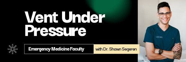
Alone With the Vent: What to Check Right After You Hook Them Up
It’s 2 a.m. There’s no RT in the building. You’ve just intubated a crashing patient, connected them to the ventilator, and for the next few minutes, it’s just you and the machine.
Initial settings matter, but what you check immediately after you hook them up is arguably more important. Here’s a framework to take to your next shift.
-
CONFIRM YOU ARE ACTUALLY VENTILATING AND OXYGENATING THE PATIENT
Before you look at the vent numbers, confirm the basics:
-
EtCO₂ present and consistent?
-
Bilateral chest rise?
-
Reasonable breath sounds?
-
Adequate SpO2?
-
CONFIRM TIDAL VOLUME (Vt) IS LUNG PROTECTIVE
Look at delivered Vt, not just what you set.
-
Target around 6 mL/kg ideal body weight in most patients
-
If Vt is unexpectedly high or low, pause and ask why
-
In pressure control modes, remember Vt will change as compliance changes
-
CHECK EXPIRATORY FLOW AND THE I:E RATIO
Now look at the waveforms.
-
Ensure expiratory flow returns to baseline before the next breath
-
Aim for an I:E ratio of at least 1:2 in most patients
-
If flow does not return to baseline, assume air trapping
This matters the most in COPD, asthma, and patients with high respiratory rates. Missing it is how hypotension shows up “out of nowhere.”
-
CONFIRM MINUTE VENTILATION MAKES SENSE
Minute ventilation is important, and it is often ignored.
-
Minute ventilation = RR × Vt
-
In most adults, a rough target is 5–10 L/min, depending on physiology
Ask yourself if this matches what the patient needs. A severe metabolic acidosis patient needs far more minute ventilation than your defaults will give them.
-
REASSESS HEMODYNAMICS
If the BP drops after starting the vent, think about too much intrathoracic pressure, air trapping, or overly aggressive settings. Improving SpO₂ at the cost of hypotension is usually not a win.
-
PLAN FOR AN EARLY BLOOD GAS
A gas within 30–60 minutes answers what the monitor cannot.
-
Is CO₂ clearance adequate?
-
Is pH acceptable for this patient?
-
Are you unintentionally worsening acidosis?
-
PAUSE AND MAKE SMALL ADJUSTMENTS EARLY
Once you have confirmed oxygenation, Vt, waveform, minute ventilation, and hemodynamics, pause and reassess. This is the moment to make small, thoughtful changes rather than waiting for alarms to force your hand.
Review this week's Vent pearls on IG.
----------
Dr. Shawn Segeren is a Canada-based Emergency Medicine physician and founder of Dynamic Simulation, a CME-accredited clinical simulation program delivering interprofessional, critical care simulations in emergency departments across Ontario.
Connect with Dr. Segeren: @drsegeren (IG) or @dynamicsimeducation (IG)
Watch the March Videos Now!
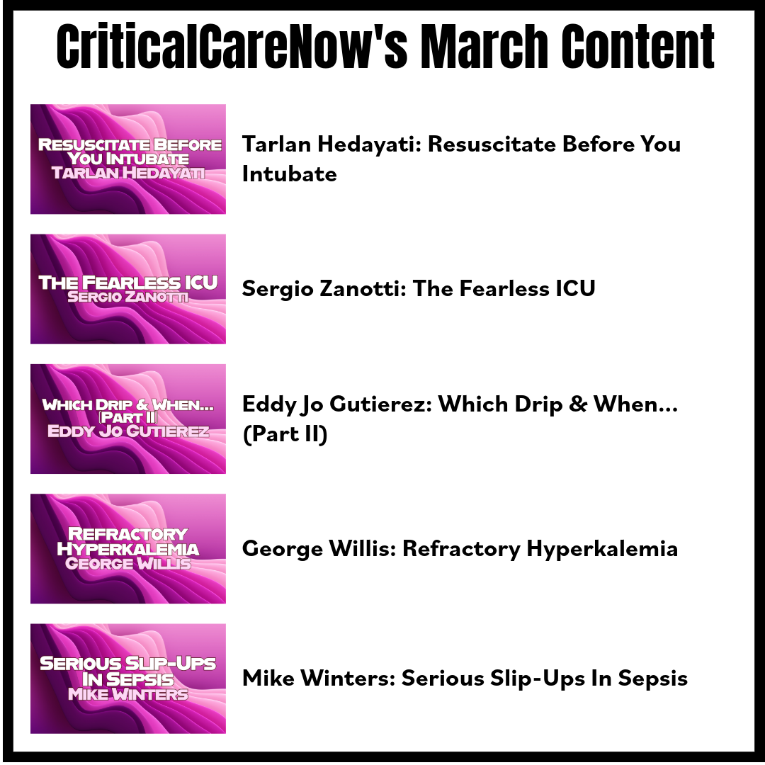
If you're an All-Access member, you're in for some great content this month. We have FIVE videos hand-picked by our staff that are high-yield and our most highly watched. We're featuring:
- Hedayati on "Resuscitate Before You Intubate"
- Zanotti on "The Fearless ICU"
- Gutierez on "Which Drip & When...(Part II)"
- Willis on "Refractory Hyperkalemia"
- Winters on "Serious Slip-Ups In Sepsis"
Each month we bring you fresh new content from the best of the best in resuscitation. If you're an All-Access member, go watch these videos NOW!


Responses