
Haney's Welcome
(Click below)

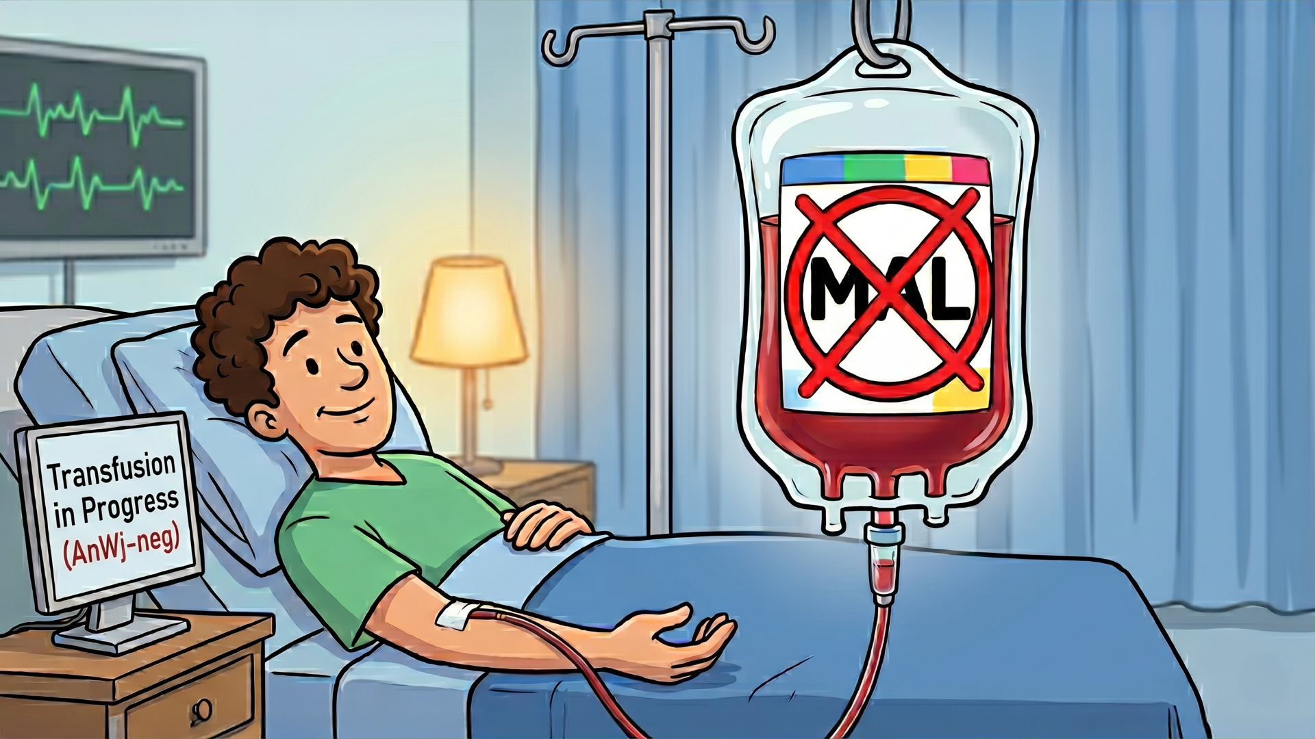
A New Blood Type Just Dropped!
Picture this: it’s 1972, a patient is pregnant, and the lab techs are absolutely losing their minds because their blood is missing a surface molecule that literally everyone else on the planet has. For 50 years, this AnWj-negative mystery was the ultimate cold case of hematology—a medical "I’ll see it when I believe it" situation that left scientists guessing. Fast forward to now, and they’ve finally cracked the code, officially christening the 47th blood group system: MAL. It turns out the missing link was a tiny, elusive protein called MAL (Myelin and Lymphocyte), which 99.9% of us are rocking on our red cells like a standard-issue uniform. If you’re in that rare 0.1% who doesn't have it, your immune system views a standard transfusion as a full-scale invasion, which is a pretty high-stakes way to find out you're "unique."
The plot twist? Being AnWj-negative isn't always something you're born with; it’s the ultimate "it’s complicated" status. While a handful of people have a literal delete-key hit on their MAL gene (shoutout to the unnamed family who helped solve this), others lose the antigen because of hematological cancers or other disorders that basically bully the protein off the cell surface. Using whole exome sequencing, researchers finally proved that the MAL gene is responsible for this antigen, ending a half-century of "we know it's there, but we can't find it." Now, instead of crossing our fingers and hoping for the best during a transfusion, we actually have the genetic receipts to identify these rare donors and patients before things get messy. It’s a massive win for precision medicine and a reminder that even after decades of clinical rounds, the human body still has a few secret levels we haven't unlocked yet.

ResusX:2026 Might Be Over, But The Education Isn't.
ResusX:2026 wrapped up last month. It was an incredible experience. Three days, live in Philadelphia. A room full of diverse clinicians who take resuscitation very seriously.
Whether you watched it live and want to revisit every moment, or you couldn't make it and need to catch up — the content is here, and it's yours for life.
Every session. Every debate. Every faculty Q&A. Coming next week, so stay tuned! In the meantime, we're giving the first 50 people who sign up below, acesss to an extra conference for FREE when they purchase ResusX:2026 Replay.
And don't forget...new ResusNation Membership tiers are coming in July 2026! More CME, more clinical depth, and more access to the education that actually moves the needle in resuscitation medicine.

Don't Sleep On IOs
Look, I'm still watching people fumble through femoral line attempts during active codes — and it's a problem. When someone is in cardiac arrest, you need access that is fast, safe for everyone in the room, and reliable enough to push anything you want into the central circulation. That's exactly what an intraosseous (IO) line gives you. IO placement takes seconds, it works, and unlike a femoral line — where I've seen people hit the artery, blow into the retroperitoneum, or spend precious minutes just finding the vein — an IO doesn't punish you or your patient for the chaos of a resuscitation.
And before someone comes at me about the single-port limitation, let's be real: you have four potential IO sites on every patient. Both tibias, both humeral heads. You can run vasopressors, push code meds, give blood — anything you'd push through a central line, you can push through an IO. There is no good reason the intraosseous line is still being underutilized in cardiac arrest. If you need reliable central access fast, stop reaching for the femoral kit and pick up the drill instead.
Watch the full video here and leave a comment.
Don't forget to like and follow my IG, TikTok, YT, Facebook or LinkedIn accounts.


Dr. George Willis delivers a masterclass warning clinicians against a lethal diagnostic pitfall: confusing decompensated hypothyroidism with standard septic shock. Because stressors like sepsis, myocardial infarctions, or strokes can trigger a precipitous metabolic decline in patients with baseline thyroid dysfunction, the true cause of death is frequently underreported on death certificates in favor of more obvious diagnoses like cardiogenic shock. Clinicians are urged to maintain a high index of suspicion and order a TSH and free T4 panel whenever evaluating an unstable, altered, or hypothermic patient whose electrolyte abnormalities or hemodynamic status fail to align with typical sepsis protocols.
Once diagnosed, managing this endocrine emergency requires avoiding several intervention traps that can worsen patient outcomes. Standard guidelines favor slow-acting T4 replacement, but Dr. Willis advocates for a low-dose combination of T3 and T4 to jumpstart metabolic drive in hypoperfused states, emphasizing that stress-dose steroids must always be administered first to prevent a catastrophic adrenal crisis. Furthermore, aggressive peripheral rewarming must be avoided in favor of simple, passive insulation to prevent worsening vasodilation and profound blood pressure drops. Finally, emergency clinicians must anticipate a highly complex, difficult airway due to the potential presence of upper airway myxedema and macroglossia, requiring expert intubation to navigate severely reduced oxygen reserves.
Check out this video of Dr. George Willis from ResusX:2026 now!


Relying on the P/F Ratio is Not Enough
The PaO2/FiO2 ratio has anchored ARDS diagnosis and severity classification for decades — it's fast, requires only an ABG and a known FiO2, and correlates with mortality at the population level. But this 2026 narrative review argues that convenience comes at a cost. The P/F ratio is a PEEP-dependent, FiO2-sensitive, hemodynamically influenced snapshot that tells you little about lung mechanics, recruitability, V/Q heterogeneity, or biological phenotype. Two patients with identical P/F ratios may face fundamentally different ventilator-induced lung injury (VILI) risk depending on driving pressure and mechanical power — a distinction the ratio cannot make. The review synthesizes complementary metrics that address these blind spots: the P/FP ratio incorporates PEEP directly and showed improved mortality discrimination across 3,400+ ARDS Network trial patients; the standardized P/F adjusts for PaCO2 to account for ventilatory failure; the ROX index captures the physiological cost of maintaining oxygenation on high-flow and remains the most validated predictor of HFNO success or failure; and the oxygenation index adds mean airway pressure for mechanistically richer context in invasively ventilated patients. None of these replaces the P/F ratio as a triage and communication tool, but each addresses a limitation in specific clinical contexts.
The deeper argument is conceptual: ARDS severity is dynamic and multidimensional. Oxygenation trajectories over the first 24–48 hours outperform baseline P/F categories for predicting ICU mortality. Hyperinflammatory biological subphenotypes — marked by elevated cytokines and endothelial injury markers — carry worse outcomes despite similar presenting P/F ratios and respond differently to corticosteroids and fluid strategy. CT analyses show that recruitable lung varies dramatically among patients in the same Berlin severity category, with direct implications for PEEP titration. The proposed framework integrates four domains — gas exchange, lung mechanics, structural and functional imaging, and molecular/immune profiling — positioning the P/F ratio not as the answer, but as the entry point into a more informed clinical evaluation.
My Takeaway Points:
- Finding - The PaO2/FiO2 ratio is highly sensitive to PEEP, FiO2, and hemodynamic status, and fails to capture lung mechanics, recruitability, or biological subphenotype — meaning identical P/F values can represent substantially different physiological states with different VILI risk profiles and treatment responsiveness.
- Practice Impact - Complementary metrics should be applied contextually: P/FP or OI for invasively ventilated patients, ROX index for those on HFNO, and stP/F when concurrent ventilatory failure is suspected — with oxygenation trajectories over 24–48 hours carrying greater prognostic weight than any single measurement.
- Population - Critically ill adults with ARDS across care settings, from resource-limited environments (Kigali modification) to ICUs managing refractory hypoxemia with prone positioning or ECMO; considerations also apply to non-intubated patients and those with high-altitude ARDS.
- Limitation - No alternative oxygenation metric has yet demonstrated consistent superiority over the P/F ratio across diverse ARDS populations or driven clear changes in therapeutic outcomes; biological subphenotyping and advanced imaging remain outside routine clinical workflows and lack guideline-level endorsement pending prospective validation.
Want to learn more? Read the full review Beyond the PaO2/FiO2 Ratio: Rethinking ARDS Severity Through the Lens of Physiology by L. Al-Husinata, et al. in Annals of Intensive Care.

Peak Pressure, Adaptive Modes, and What Actually Matters
Mechanical ventilation creates a pressure gradient to deliver flow over time — and peak airway pressure is one of the most watched numbers in the ICU because high numbers make the ventilator ding. But how much does it actually matter?
The short answer: not as much as we treat it.
Peak pressure is mostly about flow pattern if all other variables are held equal. The fundamental difference between adaptive modes like pressure-regulated volume control (PRVC) and volume-controlled ventilation with a square waveform often comes down to just a few cmH₂O, and that difference is a direct consequence of the equation of motion, not a meaningful physiological distinction. In pressure-controlled and adaptive modes, flow decelerates throughout inspiration as the pressure gradient narrows. In square-flow volume control, flow is constant, meaning resistive pressure is sustained for the entire inspiratory phase. This is why peak pressure is higher — not because the lung is under greater stress, but because the flow pattern is different.
Guldager et al. demonstrated this directly: PRVC produced a peak pressure of 20 cmH₂O versus 24 cmH₂O with square-flow volume control, with no difference in clinical outcomes. Plateau pressure which reflects the true elastic load on the lung was identical across modes. The variation in peak pressure was entirely attributable to the flow pattern and resistive load, nothing more.
It is worth noting that because normal physiologic breathing follows a decelerating flow pattern, square-flow delivery is rarely the preferred choice in clinical practice. A decelerating waveform in volume control or a pressure-targeted mode more closely approximates natural breathing mechanics.
Unlike plateau pressure and driving pressure, peak airway pressure has not been consistently associated with patient-centered outcomes in prospective studies. A difference of a few cmH₂O at the peak does not change what the alveolus experiences. Chasing a lower peak pressure is largely chasing a number.
By continuously adjusting delivered pressure to hit a target tidal volume, adaptive modes can mask evolving pathology. When respiratory mechanics worsen — bronchospasm, worsening compliance, secretions, auto-PEEP — the ventilator compensates by increasing pressure rather than alarming. This can delay recognition of a developing problem by seconds to minutes, until predefined pressure limits are finally breached. In volume control, a sudden rise in peak pressure is immediately visible and actionable. In adaptive modes, the signal is dampened by design.
There is also the issue of work of breathing. Adaptive modes cannot distinguish between changes in passive mechanics and active patient effort. In a patient who is not fully passive, the algorithm may interpret spontaneous effort as improved compliance and reduce support accordingly — potentially increasing the patient's work of breathing without any clinical warning. This interaction between the ventilator algorithm and patient effort deserves careful attention, particularly in patients who are lightly sedated or transitioning toward spontaneous breathing.
The Takeaway
Plateau pressure and driving pressure reflect what the lung actually experiences. Peak pressure reflects flow and resistance. Know the difference, choose your mode deliberately, set your alarms accordingly, and don't let a lower peak number give you false confidence that the lung is better protected.
Review this week's pearls on IG.
----------
Dr. Nicholas Ghionni (Floating Vent Guy) is a Pulmonary and Critical Care attending physician, nationally recognized vent educator, and host of The Peak Inspiration Podcast. He is passionate about translating complex ventilator mechanics and physiology into practical bedside education through engaging content, and a year-long ventilation preceptorship based out of the NIH.
Connect with Dr. Ghionni: @pulmtoilet IG / YT / Spotify / Apple
Watch the June Videos Now!

If you're an All-Access member, you're in for some great content this month. We have FIVE videos hand-picked by our staff that are high-yield and our most highly watched. We're featuring:
- Repanshek on "Crush Injuries"
- Hagahmed on "Peri-Arrest Pearls & Pitfalls"
- Mallemat & Swaminathan on "A Curious Case of Resuscitation"
- Haywood on "The Art of Pre-Oxygenation"
- Kim on "A Practical Approach to Massive Transfusion"
Each month we bring you fresh new content from the best of the best in resuscitation. If you're an All-Access member, go watch these videos NOW!


Responses